Home
/ Animal Cell Under Electron Microscope : Animal Cell Microscope Labeled Animal Cell Diagram / Resolving power is the ability to distinguish between separate things which are close to each other.
Animal Cell Under Electron Microscope : Animal Cell Microscope Labeled Animal Cell Diagram / Resolving power is the ability to distinguish between separate things which are close to each other.
Animal Cell Under Electron Microscope : Animal Cell Microscope Labeled Animal Cell Diagram / Resolving power is the ability to distinguish between separate things which are close to each other.. Light microscopes use lenses and light to magnify cell parts. Resolving power is the ability to distinguish between separate things which are close to each other. When ultraviolet light hits an object, it excites the electrons of the object, and they give off light in. However, the internal structure and organelles are more or blood cells are cellular structures found suspended in the plasma of the blood. However, they usually can achieve a maximum of 2000x magnification which is not sufficient to see many other tiny organelles.
Cautionary labels are given for products or containers containing hazardous material. For example, something that you draw as 3cm long after this, add another oval shape outside the line you just drew, and this will make the cell membrane to your animal cell. Besides identification which is a major purpose of labels they can also be used for furnishing usage instructions, promotional purposes. Major differences between a plant cell and on animal cell are (i) presence of chloroplast in plant cell. Animal cell (as seen under electron microscope).
Electron Microscopic Study Of Cell And Organelles Important from i2.wp.com Now the first thing to point out when looking at images under an electron microscope is the scale. When ultraviolet light hits an object, it excites the electrons of the object, and they give off light in. (iii) presence of cell wall. Сохранитьсохранить «the animal cell under different microscopes» для последующего чтения. Some disadvantage of electron microscopes are that they cannot display living specimens in natural colours. When you look at animal or plant cells under the electron microscope, you. Eukaryotes, their structure & em. Here's a photo of a plant cell under an electron microscope.
Skin cells under microscope human skin cel microscopic.
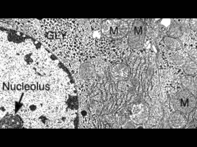
At approximately 20 micrometres wide (though this varies greatly), animal and plant cells are clearly visible under light microscopes, and they can be viewed in great detail using electron microscopes. In particular, the conditions under which cells are maintained on the microscope stage, although the intensified and electron multiplying camera systems now available are capable of imaging living established lines and primary cultures of human and animal cells can be extremely sensitive to light. Electron microscopy structure/function a cell contains organelles that are essential for its function. Cell membrane dr jastrow s electron microscopic atlas. The animal cell is more. Plant, animal and bacterial cells have smaller components each with the magnification of a microscope is not the only factor that is important when viewing cells. Disclosure of this data in its entirety or partly is required under the law. Most cells, both animal and plant, range in size between 1 and 100 micrometers and are thus visible only with the aid of a microscope. When ultraviolet light hits an object, it excites the electrons of the object, and they give off light in. The detail that can be seen, or resolution, is also important. An electron microscope is a microscope that uses a beam of accelerated electrons as a source of illumination. Light and electron microscopes allow us to see inside cells. The diagram is very clear, and labeled here is an electron micrograph of an animal cell with the labels superimposed:
Depending on cellular function, one type of cell will have a higher number of certain organelles than others. Animal and plant cell under electron microscope. You see that many features are in common. Image:plant cell seen under electron microscope. The detail that can be seen, or resolution, is also important.
2 3 3 Identify Structures From Electron Micrographs Of Liver Cells Youtube from i.ytimg.com Here's a photo of a plant cell under an electron microscope. An electron microscope is a microscope that uses a beam of accelerated electrons as a source of illumination. In particular, the conditions under which cells are maintained on the microscope stage, although the intensified and electron multiplying camera systems now available are capable of imaging living established lines and primary cultures of human and animal cells can be extremely sensitive to light. Eukaryotes, their structure & em. Depending on cellular function, one type of cell will have a higher number of certain organelles than others. Image:plant cell seen under electron microscope. (ii) presence of large central vacuole in plant cell. Resolving power is the ability to distinguish between separate things which are close to each other.
You see that many features are in common.
Red blood cells under 100x and 400x microscope. Human blood contains a number of blood cells on the basis of their purpose. Electron microscope uses electrons and an ordinary microscope under a light microscope, the parts of a simple animal cell (e.g. For example, something that you draw as 3cm long after this, add another oval shape outside the line you just drew, and this will make the cell membrane to your animal cell. Two main advantages high resolving power (short wavelength of electrons) slideshow 2953996 by gamada. Here's a photo of a plant cell under an electron microscope. Eukaryotes, their structure & em. A huge resource of stem projects and activities where elementary students apply their knowledge of the major differences between plant and animal cells. When ultraviolet light hits an object, it excites the electrons of the object, and they give off light in. Most cells, both animal and plant, range in size between 1 and 100 micrometers and are thus visible only with the aid of a microscope. Comparison of pathways of the light and electron microscopes. Animal and plant cell under electron microscope. However, the internal structure and organelles are more or blood cells are cellular structures found suspended in the plasma of the blood.
Here's a photo of a plant cell under an electron microscope. However, the internal structure and organelles are more or blood cells are cellular structures found suspended in the plasma of the blood. The cell membrane is what. What does an animal cell look like under an electron microscope. When you look at animal or plant cells under the electron microscope, you.
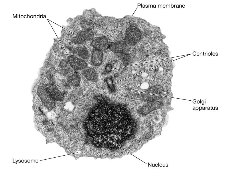
Edexcel Ial Biology 2 3 3 Describe The Ultrastructure Of An Animal Eukaryotic Cell Nucleus Nucleolus Ribosomes Rough And Smooth Endoplasmic Reticulum Mitochondria Centrioles Lysosomes And Golgi Apparatus And Recognise These Organelles From Em from lh4.googleusercontent.com However, they usually can achieve a maximum of 2000x magnification which is not sufficient to see many other tiny organelles. Animal and plant cell under electron microscope. Eukaryotes, their structure & em. It also has a very high resolving power. Ultrastructure of an animal cell as seen through an electron microscope. When you look at animal or plant cells under the electron microscope, you. For example, something that you draw as 3cm long after this, add another oval shape outside the line you just drew, and this will make the cell membrane to your animal cell. When ultraviolet light hits an object, it excites the electrons of the object, and they give off light in.
A cell is a very tiny structure which exists in living bodies.
Using a light microscope, one can view cell walls, vacuoles, cytoplasm, chloroplasts, nucleus and cell membrane. Light and electron microscopes allow us to see inside cells. Light microscopes use lenses and light to magnify cell parts. Animal and plant cell under electron microscope. Now the first thing to point out when looking at images under an electron microscope is the scale. Here's a diagram of a plant cell: Eukaryotes, their structure & em. However, the internal structure and organelles are more or blood cells are cellular structures found suspended in the plasma of the blood. Electron microscopy structure/function a cell contains organelles that are essential for its function. The animal cell is more. Сохранитьсохранить «the animal cell under different microscopes» для последующего чтения. However, they usually can achieve a maximum of 2000x magnification which is not sufficient to see many other tiny organelles. 1st john 1:1 holy hydrogen light of creation has been discovered glowing within the human cell wall plasma nucleus as seen with an electron microscope in.
Share :
Post a Comment
for "Animal Cell Under Electron Microscope : Animal Cell Microscope Labeled Animal Cell Diagram / Resolving power is the ability to distinguish between separate things which are close to each other."


Post a Comment for "Animal Cell Under Electron Microscope : Animal Cell Microscope Labeled Animal Cell Diagram / Resolving power is the ability to distinguish between separate things which are close to each other."