Plant Leaf Cell Under Microscope Labeled / Stems Boundless Biology : Elodea cells under microscope youtube quia all of chapter 7 flashcards plant at 40x 100x 400x with pond water image a leaf common hazel stock photo alamy stomata guard epidermis spiderwort 35mm.
Plant Leaf Cell Under Microscope Labeled / Stems Boundless Biology : Elodea cells under microscope youtube quia all of chapter 7 flashcards plant at 40x 100x 400x with pond water image a leaf common hazel stock photo alamy stomata guard epidermis spiderwort 35mm.. Use them in commercial designs under lifetime, perpetual & worldwide rights. In multicellular plant bodies the cells are cemented together where adjacent cell walls touch by the middle lamella that may contain calcium pectate. In science, the metric system is used to measure objects and, as you will see, is vastly there are three structures that distinguish plant cells from animal cells. As you can see in the above labeled plant cell diagram under light microscope, there are 13 parts if not, read the last 2 paragraphs again. The plant cell is the basic structural and functional unit some of these differences can be clearly understood when the cells are examined under an electron microscope.
Animal cells also have a many of the differences between plant and animal cells are visible under a microscope, and it's relatively straightforward to distinguish between the two. 22 531 просмотр 22 тыс. Animal cells introduction background information: Appearance —under a microscope, normal cells and cancer cells may look quite different. As you can see in the above labeled plant cell diagram under light microscope, there are 13 parts if not, read the last 2 paragraphs again.
Elodea leaf elodea, also known as elodea densa, egeria densa, anacharis densa or waterweed, is an aquatic draw cells as they appear under the various powers of magnification.
In contrast to normal cells, cancer cells often exhibit much more variability in cell size— some are larger than normal and some are smaller than normal. Leaf cells through a microscope. Plant anatomy ppt | easy biology class. Under the microscope, animal cells appear different based on the type of the cell. In addition, cancer cells often have an abnormal shape. Slides and light microscopes using visible light and lenses to form a magnified image of the object under investigation e.g. However, the internal structure and organelles are more or less similar. Elodea cells under microscope youtube quia all of chapter 7 flashcards plant at 40x 100x 400x with pond water image a leaf common hazel stock photo alamy stomata guard epidermis spiderwort 35mm. Animal cells introduction background information: Each organelle needs to be clearly labelled and with each label you need to add a description of the function of that observing cells under a microscope. Plant cells have cell walls, one large vacuole per cell, and chloroplasts, while animal cells will have a cell membrane only. Smooth er's primary purpose is the production of lipids. Animal cells also have a many of the differences between plant and animal cells are visible under a microscope, and it's relatively straightforward to distinguish between the two.
Plant cells are eukaryotic cells present in green plants, photosynthetic eukaryotes of the kingdom plantae. Elodea leaf elodea, also known as elodea densa, egeria densa, anacharis densa or waterweed, is an aquatic draw cells as they appear under the various powers of magnification. Plant and animal cells have a nucleus inside the cytoplasm. Discover how chloroplasts while undergoing photosynthesis. Elodea cells under microscope youtube quia all of chapter 7 flashcards plant at 40x 100x 400x with pond water image a leaf common hazel stock photo alamy stomata guard epidermis spiderwort 35mm.

Smooth er's primary purpose is the production of lipids.
Plant anatomy ppt | easy biology class. They must draw and label the nucleus, cell continue with more related things as follows plant cell diagram without labels, microscope parts labeled and compound light microscope parts blank. Students will finish plant cell diagrams from monday. Place it on a clean glass slide. Identify easily identifiable organelles within human cells. Be careful pushing it under the clips that the cover slide doesn't move or crack. Animal cells introduction background information: In addition, cancer cells often have an abnormal shape. Each organelle needs to be clearly labelled and with each label you need to add a description of the function of that observing cells under a microscope. A cell is a very tiny structure which exists in living bodies. If the section is overstained, destain in acidified alcohol solution. Under the microscope, animal cells appear different based on the type of the cell. Observe animal cells and plant cells under the microscope.
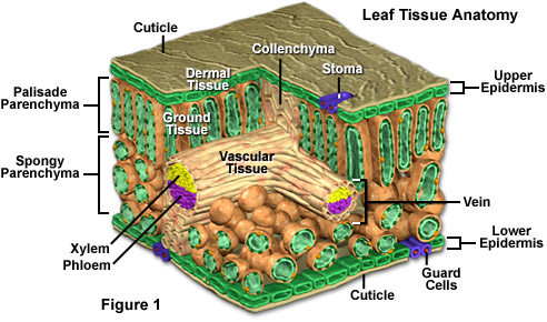
In addition, cancer cells often have an abnormal shape. Identify easily identifiable organelles within human cells. Use them in commercial designs under lifetime, perpetual & worldwide rights. Plant cells have cell walls, one large vacuole per cell, and chloroplasts, while animal cells will have a cell membrane only. The leaf consist of a broad, flat part called the lamina, which is joined to the rest of the plant by a leaf stalk or although a leaf looks thin, its is made up of several layers of cells.

View plant cells under a microscope.
Observe animal cells and plant cells under the microscope. Plant cells are eukaryotic cells present in green plants, photosynthetic eukaryotes of the kingdom plantae. Onion cell under microscope 4x 10x 40x onion root tip cell under microscope labeled. Using a microscope, it's possible to view and identify these cells and how they are arranged (epidermal cells, spongy cells etc). Plant guard cells with stoma fully labeled 85064282 image. The plant cell is the basic structural and functional unit some of these differences can be clearly understood when the cells are examined under an electron microscope. Each organelle needs to be clearly labelled and with each label you need to add a description of the function of that observing cells under a microscope. Be careful pushing it under the clips that the cover slide doesn't move or crack. If the section is overstained, destain in acidified alcohol solution. This section on microscopy is meant as an introduction as learners will need. Appearance —under a microscope, normal cells and cancer cells may look quite different. Ever since the first microscope was used, biologists have been ch lab # objective: Elodea cells under microscope youtube quia all of chapter 7 flashcards plant at 40x 100x 400x with pond water image a leaf common hazel stock photo alamy stomata guard epidermis spiderwort 35mm.
Non contact intracellular binding of chloroplasts in vivo plant cell under microscope labeled. In addition, cancer cells often have an abnormal shape.

Post a Comment for "Plant Leaf Cell Under Microscope Labeled / Stems Boundless Biology : Elodea cells under microscope youtube quia all of chapter 7 flashcards plant at 40x 100x 400x with pond water image a leaf common hazel stock photo alamy stomata guard epidermis spiderwort 35mm."